Successful application of BONITex® implants in dental rehabilitation procedure
Summary
This case study demonstrates the successful use of BONITex® implants in complex dental rehabilitation, involving tooth extraction, implantation, augmentation, and soft tissue correction. The procedure resulted in improved oral function and aesthetics for the patient.
TELESCOPIC HYBRID BRIDGE
Implant prosthesis after maxillary cleft osteoplasty
by: DR. INGO BUTTCHEREIT, Rolf H. YTREHUS, PD Dr. Dr. PEER W. KÄMMERER
The oral rehabilitation of patients with cleft lip, jaw, and palate requires thorough planning. This involves conducting a systematic survey that includes assessing the dental and periodontal status, functional findings, model analysis, and analyzing the anatomical situation. Attention must be given to alveolar process defects, scarring, residual palate perforations, premaxilla mobility, and physiognomy.
In the case of an unfavorable anatomical or dental/periodontal initial
situation, the removable telescoping bridge represents a proven treatment concept. The removable bridge combines the advantages of fixed and removable
dentures. Conventional bridge restorations without augmentative procedures are seldom suitable for aesthetic reasons and hygiene, particularly when dealing with vertical bone deficits.
Case Study
In March 2017, a 45-year-old patient visited the Department of Oral and Maxillofacial Surgery at the University of Rostock for implant surgery. The patient had previously undergone a soft tissue operation for a left cleft lip and palate. The family dentist sought advice regarding the possibility of implant-supported treatment for teeth 21-24.

Intraorally, there was a technically intact but aesthetically inadequate fixed restoration in teeth 11-23, which required frequent re-cementation due to loosening. Tooth 11 and 23 exhibited visible crown edges with suspected secondary caries. Radiological assessments (Fig. 1a, 1b) revealed a bony deficit in tooth 21/22, measuring approximately 4×2 cm, indicating a complete jaw cleft, along with vertical bone collapses distally on tooth 11 and mesially on tooth 23. Additionally, there was apical osteolysis and insufficient root canal treatment in tooth 23 and tooth 11.
Based on the initial findings, the treatment plan included jaw gap osteoplasty in the region of 21/22, implant insertion in the regions of 11/21, 23, and 25, and the implementation of an implant-supported bridge. The jaw gap osteoplasty and the removal of non-viable teeth 11 and 23 were performed in March 2017 during the patient's inpatient stay under intubation anesthesia.
Intraoperatively, after the extraction of teeth 11 and 23 (Fig. 2) through a palatal incision (with vertical relief incisions in the vestibular regions 12 and 26), mucoperiosteal flaps were created both palatally and vestibularly, revealing the bone defect indicative of a complete jaw cleft in the 21/22 region (Fig. 3). The defect was reconstructed using autologous bone obtained from the left iliac crest (bone punches and cortical bone; (Fig. 4) through lateral and vertical augmentation techniques, along with the jaw gap osteoplasty. Postoperative mobilization progressed smoothly under physiotherapeutic supervision, allowing for pain-adapted full weight bearing. Due to recurring swelling and intermittent fever episodes, the patient's hospital stay was extended, and she was discharged on the 6th day after surgery.
For patients without a cleft lip and palate, the prosthetics can be planned with careful consideration of physiology, stability, aesthetics, hygiene, and the patient's expectations.

After the wound had healed, tooth 27, which was not worth preserving, was extracted under local anesthesia at the end of April 2017. A follow-up appointment was scheduled in December 2017 to plan the actual implantation. In this context, a dental volume tomography (DVT) was performed, revealing partial resorption of the previous osteoplasty. As a result, implantation in region 11/21 appeared unfeasible without additional augmentation. Consequently, implant planning was revised for regions 22 and 24, and a conversion of the prosthetic concept was carried out. The revised plan included a telescopic bridge on the natural pillars 12 and 26, as well as the planned implants 22 and 24.
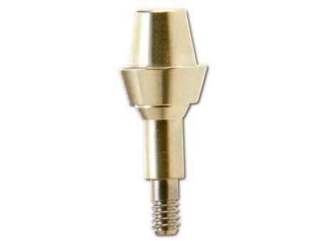
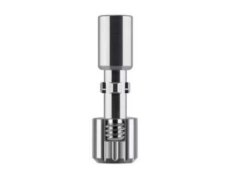
The successful outcome of the surgery was attributed to the utilization of CTD-alphatech Tube-Line BONITex® implants.
After thoroughly explaining the changes to the patient, guided insertion of two CTD-alphatech Tube-Line BONITex® implants was performed in regions 22 (3.8×10 mm) and 24 (4.3×10 mm) using a 3D drilling template and local anesthesia.
Following a tertiary jaw gap osteoplasty and implantation in regions 22 and 24, the inadequate soft tissue condition in the vestibulum region 12 to 26 was corrected in April 2018 under intubation anesthesia. This was achieved by performing a vestibuloplasty using buccal mucosa. The non-keratinized gingiva was mobilized cranially and basally on the periosteum with resorbable suture material (Vicryl 4.0) and fixed in the area of the nasal floor with non-resorbable suture material (Resolon 4.0). Cheek mucosa was also removed from the right cheek, and the transplant was fixed with non-resorbable cash suture material (Resolon 4.0) in the vestibular region 12-25. An individual bandage plate lined with Aqueron (Erkodent Erich Kopp GmbH, Pfalzgrafenweiler, Germany) was incorporated.
Ten days after surgery, the bandage plate was removed and necessary sutures were adjusted. Eight weeks after the vestibuloplasty, the implants in regions 22 and 24 were exposed using a rolled flap, and the surgical treatment was completed with proper suturing. Throughout the surgical treatment, the patient wore an interim maxillary prosthesis. Subsequently, the family dentist (ZA Dr. med. dent. Harald Riemer/Rostock) initiated the fabrication of a telescoping hybrid bridge from 12 to 26 in the upper jaw. (Figs. 10-12).
The 2-year follow-up revealed consistently stable peri-implant conditions with no signs of pathological probing depths. Radiologically, the follow-up results were also unremarkable, indicating successful long-term outcomes. (Fig. 13)
Discussion
Osteoplasty in cleft patients can be performed primarily during infant dentition, secondarily during mixed dentition, or tertiary during permanent dentition. There is no established standard that universally recommends the timing of the surgery, the choice of materials, or the surgical technique. Likewise, there is no standardized and objective evaluation criterion for measuring success [9].
However, clinically, secondary osteoplasty using iliac crest spongiosa during the early phase of mixed dentition (between the ages of 7 and 11 years) has shown positive results. In two comparative studies conducted in 2002, secondary osteoplasty demonstrated slight advantages over tertiary osteoplasty in terms of postoperative resorption [4, 10]. Due to the size of the defect, it is typically necessary to transplant bone from an extraoral source, often from the ridge itself.
This procedure of harvesting bone carries certain risks. Known complications include persistent pain, nerve injuries, bleeding, changes in gait, scarring, bone alterations, infections, fractures, peritonitis, and even the formation of abscesses [3]. Alternatively, bone can be obtained from the fibula, tibia, or lower jaw. Nowadays, allogeneic replacement materials can also be utilized to avoid the need for an extraoral donor site. However, it is important to consider complications such as osteoclast activation and wound healing disorders when using collagen soaked with bone morphogenic protein [2]. The use of allogeneic or xenogeneic alternatives should not overlook the lack of clinical evidence, cost factors, and material availability.
The key to achieving long-term success is the earliest possible loading (3-6 months) after the operation. Loading can be achieved through normal tooth eruption (after secondary osteoplasty) or with the use of an implant (after tertiary osteoplasty). Implantation in the upper jaw can be conventionally planned by assessing the available bone after osteoplasty.
A clinical study conducted from 1994 to 1998 involving 15 patients and a total of 23 implants treated with an iliac crest transplant in the gap area after osteoplasty showed an implant survival rate of 96% after 2.5 years [6]. A similar study in 2020 reported a 95% implant survival rate after implantation in the gap area [1].
In summary, it has been demonstrated that the success rates of implantation in the gap area are comparable to those in native, non-transplanted bone. The results regarding peri-implant soft tissue and aesthetics are also considered highly satisfactory [1]. However, a portion of the study participants (18/40; 45%) required re-augmentation ("replacement osteoplasty") during implantation due to postoperative resorption [1, 6].
After the completion of surgical therapy, prosthetic care is necessary to restore function, phonetics, and aesthetics. The prosthetic treatment can be planned similarly to patients without a cleft lip and palate, considering factors such as physiology, stability, aesthetics, hygiene, and patient expectations [5]. The restoration can involve the use of natural teeth, implants, or a combination of both. One important consideration for many patients is the impression-taking process. Due to the potential presence of an oronasal fistula, it is important to use an impression material that is not overly viscous, as it may be difficult to remove from the impression tray and cause additional discomfort for the patient. If a fistulous tract is still present, it should be covered before taking the impression, taking into account its size [8].
Regarding prosthetic constructions, a review conducted in 2012 examined 25 included studies on hybrid prosthetics. The review reported an implant survival rate of 75-100%, a dental complication rate of 5.4-11.8%, intrusion with removable superstructures ranging from 0-66%, and intrusion with fixed supra-structures ranging from 0-44%. Overall, removable constructions appear to exert less mechanical stress on the superstructure, while teeth and implants experience greater stress. However, fixed abutments tend to yield more favorable clinical outcomes due to reduced intrusion of natural teeth [7].
Conflicts of interest: The authors, Dr. Ingo Buttchereit and Rolf H. Ytrehus, declare no conflicts of interest related to this contribution or outside of the submitted work. The author, PD Dr. Dr. Peer W. Kämmerer, also states no conflicts of interest in connection with the submitted work. Outside of the submitted work, Dr. Kämmerer reports financial activities involving Knowledge Compact, ZZI, Sanofi-Aventis, Straumann, Dental Association, ITI, and Zimmer Biomet.
Conclusion For Practice
References

Author: DR. INGO BUTTCHEREIT, Rolf H. YTREHUS, PD Dr. Dr. PEER W. KÄMMERER


